Utilizing Pinch Anatomy for PDO Threads
What Is Pinch Anatomy?
What is
pinch anatomy and how does it apply to PDO threads?
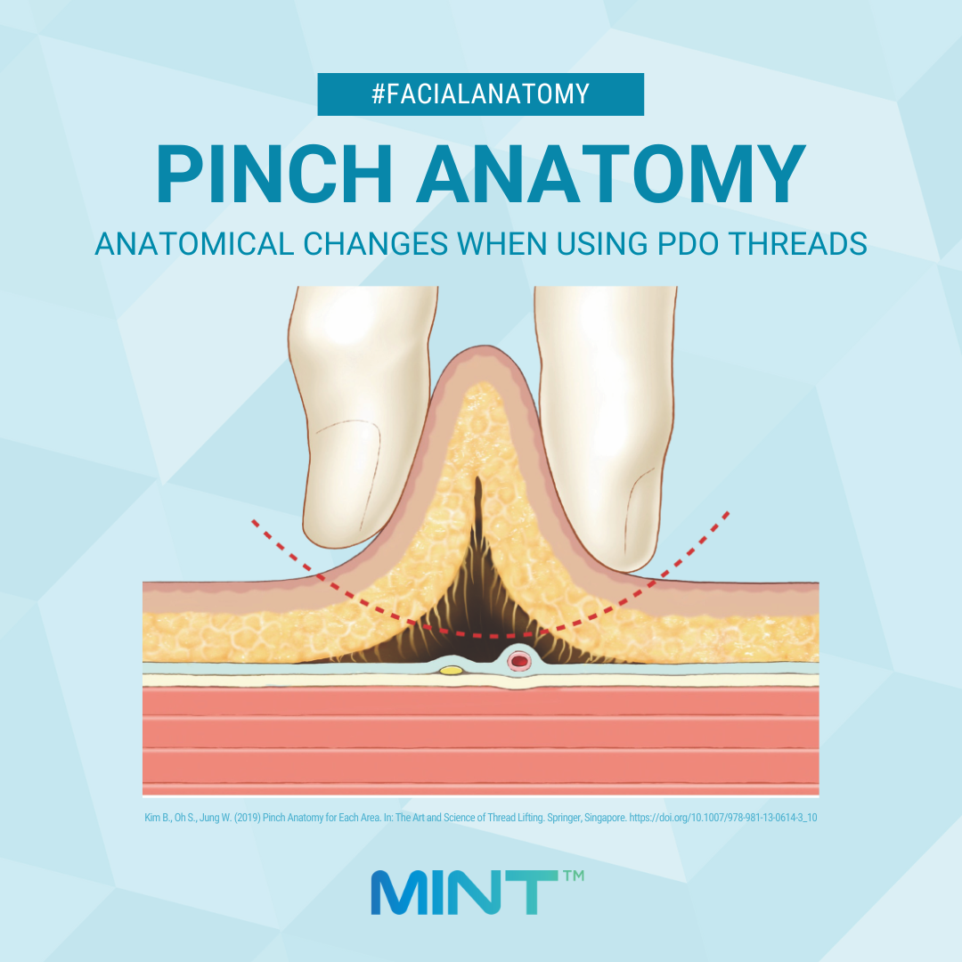
Medical illustrations or cadaver dissections generally show facial structures without changes in their anatomical structures. However, practitioners often pinch or pull the skin during patient assessment and treatment for PDO threads, which changes its original appearance. This
difference in the internal tissue through pinching or pulling is referred to as pinch anatomy.
When you pinch the tissues, the underlying blood vessels and nerves move with them. What layer of the skin and whether blood vessels and nerves are pulled upward during this process can determine if going deeper or more superficial would be the safer treatment method.
Knowing what anatomical changes occur through different methods of pinching can help minimize the risk of complications, such as blood vessel damage in the temporal area or nerve damage in the zygomatic arch area.
You can read
"Pinch Anatomy for Each Area. In: The Art and Science of Thread Lifting" by Bongcheol Kim, Seungmin Oh, and Wonsung Jung to learn more about pinch anatomy and areas where it is important.
To put things you have learned into practice, join us for one of our hands-on training sessions. For upcoming dates, please click here.
References
Kim B., Oh S., Jung W. (2019) Pinch Anatomy for Each Area. In: The Art and Science of Thread Lifting. Springer, Singapore. https://doi.org/10.1007/978-981-13-0614-3_10